- MR Cardiac Suite
-
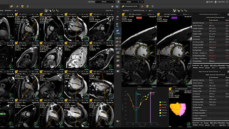
An enhanced cardiac MR reading experience
MR Cardiac Suite debuts a new, award-winning look and feel³, enabling seamless review and analysis of cardiac MR studies in one comprehensive environment. Enhanced reading experience with multimodality 2D & 3D viewing, comparison to priors and flexible and personalized layouts. Highly intuitive environment, enabling users to complete the majority of tasks with minimal training⁴. - Enhanced cardiac workflow
-
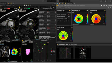
Results-driven and personalized Cardiac MR workflow
An enhanced workflow to processing cardiac MR studies including automatic LV/RV contouring, user-defined viewing protocols and customization according to the clinical questions⁵, the user can also set their own normal values. It also includes consolidated findings dashboard with all results in one place. - Cardiac analysis
-
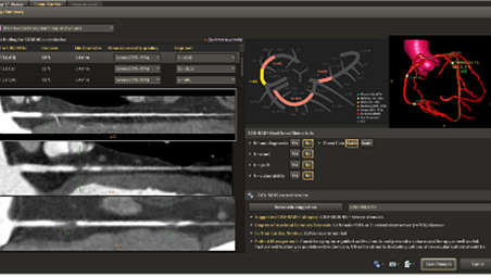
Comprehensive cardiac analysis with CAD-RADS functionality
Integration of coronary lesions in a single view to support determination of disease severity and results communication. Findings management with all findings in one screen, with various layouts and views. Semi-automatic CAD-RADS workflow embedded within the application, the functionality enables standardized communication of results. The multi-batch option enables saving MPR images of all selected coronaries at once, to the PACS or any other configured devices. - Ischemic stroke case support
-
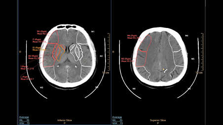
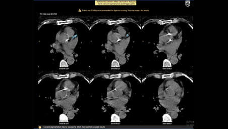
CT ASPECT Scoring¹,² supporting ischemic stroke cases
Supports in the review and analysis of non-contrast CT images for the management of ischemic stroke patients with a known large Vessel Occlusion (LVO) such as a Mid Cerebral Artery (MCA) or Internal Carotid Artery (ICA) occlusion with an automated workflow. The application automatically registers images, segment regions and provides calculation of ASPECT score and auto-send results to PACS. - LOBI
-
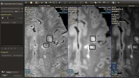
MR Longitudinal Brain Analysis (LOBI)
The MR Longitudinal Brain Analysis highlights subtle neurological changes over time. The application now supports the generation of FLAIR* series. The FLAIR* could aid in the visualization of central vein sign in white matter lesions, which may help with the diagnosis of multiple sclerosis6. Now you can view FLAIR* map, compare all 3 planes of all series and save FLAIR* as new DICOM series. - Advanced vessel analysis
-
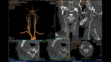
Advanced vessel analysis with spectral results
A single application for both conventional and spectral CT data with improved workflow and user experience, designed for faster results. The application supports all spectral results for comprehensive vessel analysis. Use Spectral Magic Glass for side-by-side review of multiple spectral results. Photo Realistic Volume Rendering is now available for spectral data. - Coronary calcified plaques
-
CT Calcium Automated Analysis¹
CT Calcium Automated Analysis¹ provides information about coronary calcified plaques on non-gated, non-contrast chest CTs, based on the SCCT 2016 guidelines. The application offers you key-images with segmented calcium for inspection, besides coronary calcium category scale and patient management recommendations. The application allows automatic results being sent straight to PACS.
An enhanced cardiac MR reading experience
An enhanced cardiac MR reading experience
An enhanced cardiac MR reading experience
Results-driven and personalized Cardiac MR workflow
Results-driven and personalized Cardiac MR workflow
Results-driven and personalized Cardiac MR workflow
Comprehensive cardiac analysis with CAD-RADS functionality
Comprehensive cardiac analysis with CAD-RADS functionality
Comprehensive cardiac analysis with CAD-RADS functionality
CT ASPECT Scoring¹,² supporting ischemic stroke cases
CT ASPECT Scoring¹,² supporting ischemic stroke cases
CT ASPECT Scoring¹,² supporting ischemic stroke cases
MR Longitudinal Brain Analysis (LOBI)
MR Longitudinal Brain Analysis (LOBI)
MR Longitudinal Brain Analysis (LOBI)
Advanced vessel analysis with spectral results
Advanced vessel analysis with spectral results
Advanced vessel analysis with spectral results
CT Calcium Automated Analysis¹
CT Calcium Automated Analysis¹
CT Calcium Automated Analysis¹
- MR Cardiac Suite
- Enhanced cardiac workflow
- Cardiac analysis
- Ischemic stroke case support
- MR Cardiac Suite
-
An enhanced cardiac MR reading experience
MR Cardiac Suite debuts a new, award-winning look and feel³, enabling seamless review and analysis of cardiac MR studies in one comprehensive environment. Enhanced reading experience with multimodality 2D & 3D viewing, comparison to priors and flexible and personalized layouts. Highly intuitive environment, enabling users to complete the majority of tasks with minimal training⁴. - Enhanced cardiac workflow
-
Results-driven and personalized Cardiac MR workflow
An enhanced workflow to processing cardiac MR studies including automatic LV/RV contouring, user-defined viewing protocols and customization according to the clinical questions⁵, the user can also set their own normal values. It also includes consolidated findings dashboard with all results in one place. - Cardiac analysis
-
Comprehensive cardiac analysis with CAD-RADS functionality
Integration of coronary lesions in a single view to support determination of disease severity and results communication. Findings management with all findings in one screen, with various layouts and views. Semi-automatic CAD-RADS workflow embedded within the application, the functionality enables standardized communication of results. The multi-batch option enables saving MPR images of all selected coronaries at once, to the PACS or any other configured devices. - Ischemic stroke case support
-
CT ASPECT Scoring¹,² supporting ischemic stroke cases
Supports in the review and analysis of non-contrast CT images for the management of ischemic stroke patients with a known large Vessel Occlusion (LVO) such as a Mid Cerebral Artery (MCA) or Internal Carotid Artery (ICA) occlusion with an automated workflow. The application automatically registers images, segment regions and provides calculation of ASPECT score and auto-send results to PACS. - LOBI
-
MR Longitudinal Brain Analysis (LOBI)
The MR Longitudinal Brain Analysis highlights subtle neurological changes over time. The application now supports the generation of FLAIR* series. The FLAIR* could aid in the visualization of central vein sign in white matter lesions, which may help with the diagnosis of multiple sclerosis6. Now you can view FLAIR* map, compare all 3 planes of all series and save FLAIR* as new DICOM series. - Advanced vessel analysis
-
Advanced vessel analysis with spectral results
A single application for both conventional and spectral CT data with improved workflow and user experience, designed for faster results. The application supports all spectral results for comprehensive vessel analysis. Use Spectral Magic Glass for side-by-side review of multiple spectral results. Photo Realistic Volume Rendering is now available for spectral data. - Coronary calcified plaques
-
CT Calcium Automated Analysis¹
CT Calcium Automated Analysis¹ provides information about coronary calcified plaques on non-gated, non-contrast chest CTs, based on the SCCT 2016 guidelines. The application offers you key-images with segmented calcium for inspection, besides coronary calcium category scale and patient management recommendations. The application allows automatic results being sent straight to PACS.
An enhanced cardiac MR reading experience
An enhanced cardiac MR reading experience
An enhanced cardiac MR reading experience
Results-driven and personalized Cardiac MR workflow
Results-driven and personalized Cardiac MR workflow
Results-driven and personalized Cardiac MR workflow
Comprehensive cardiac analysis with CAD-RADS functionality
Comprehensive cardiac analysis with CAD-RADS functionality
Comprehensive cardiac analysis with CAD-RADS functionality
CT ASPECT Scoring¹,² supporting ischemic stroke cases
CT ASPECT Scoring¹,² supporting ischemic stroke cases
CT ASPECT Scoring¹,² supporting ischemic stroke cases
MR Longitudinal Brain Analysis (LOBI)
MR Longitudinal Brain Analysis (LOBI)
MR Longitudinal Brain Analysis (LOBI)
Advanced vessel analysis with spectral results
Advanced vessel analysis with spectral results
Advanced vessel analysis with spectral results
CT Calcium Automated Analysis¹
CT Calcium Automated Analysis¹
CT Calcium Automated Analysis¹
Related products
Alternative products

- ¹ Not available in the US
- ² These functionalities may not be available in all territories. Please contact Philips representative for more details.
- ³ IF Design Award, 2022
- ⁴ More than 90% of frequently performed tasks completed independently, with max 1.5 hr training for the entire MR Cardiac suite.
- ⁵ Common clinical questions: Ischemic heart disease and Non-ischemic cardiomyopathy (with LV & RV functional analysis, T1/T2/T2* Mapping, Stress-rest perfusion, and Late Gadolinium Enhancement).
- ⁶ Sati P et al. Nat Rev Neurol. 2016 ;12(12):714-722
- Product not available for sale in all countries. Please contact your sales representative to determine availability in your country.