Heidelberg University Hospital adds diagnostic value with spectral imaging
Customer story ∙ By Philips Healthcare ∙ Featuring Heidelberg University Hospital, Heidelberg, Germany ∙ Φεβ 14, 2023 ∙ 4 min read
As an interdisciplinary hub, Heidelberg University Hospital depends on highly precise diagnostics for complex cases, requiring imaging solutions that allow for quick and reliable assessment, particularly for cardiology and oncology patients. Philips Spectral CT 7500 features “always-on” dual-layer spectral-detector technology that offers spectral results in a single scan, available even retrospectively. The 8 cm detector improves cardiac imaging, including for patients with high or irregular heart rates. Spectral reconstruction and analysis are seamlessly integrated into the existing workflow.
Customer story at a glance
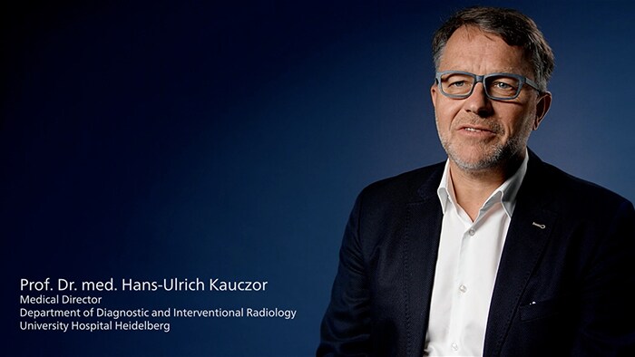
Prof. Dr. Hans-Ulrich Kauczor describes the experience of Heidelberg University Hospital with Philips Spectral CT 7500.
Spectral-detector imaging is an investment that creates both clinical and economic value
Heidelberg University Hospital has more than 2,000 beds and treats more than 80,000 inpatients and more than a million outpatients a year, focusing on oncology, cardiovascular disease, and chronic and acute inflammatory diseases.
“We see more. And what we may have seen before, we can now say what it is with greater certainty.”
Prof. Dr. Hans-Ulrich Kauczor
Prof. Dr. Hans-Ulrich Kauczor describes the value of spectral technology with dual-layer NanoPanel Prism detectors that absorb the low- and high-energy photons to automatically capture and archive spectral image information with each scan. “Spectral imaging creates clear added value in diagnostics,” he says. “We see more. And what we may have seen before, we can now say what it is with greater certainty. We also have the complete spectral information available.”
"Spectral-detector CT is a fast diagnostic tool. Studies show that scans with detector-based spectral CT help physicians achieve 34% shorter time to diagnosis compared to conventional CT scans in certain cases [1]. This is in line with the experience of the Heidelberg team.
"As clinic director, I have to make sure that I not only achieve clinical but also economic added value," says Kauczor. “I can say that this investment pays off. With Philips Spectral CT 7500 we can achieve both.” Spectral is fast. Examinations of the thorax and head take less than a second, and a CT scan of the entire upper body takes less than two seconds. In addition, the new 100 kV scan mode offers great potential for saving radiation dose.
Delivering more sensitive results in oncology
Rapid and sensitive diagnostics are essential for the diagnosis, treatment and follow-up of cancer patients. Prof. Dr. Dirk Jaeger talks about the major increase in the quality of oncology imaging made possible by spectral-detector imaging. "With Spectral CT 7500, we are significantly more sensitive when it comes to detecting lesions and detection reliability,” he says. He notes that metastases can be diagnosed more reliably and recurrences detected much earlier. "It's a blessing for patients," he concludes.
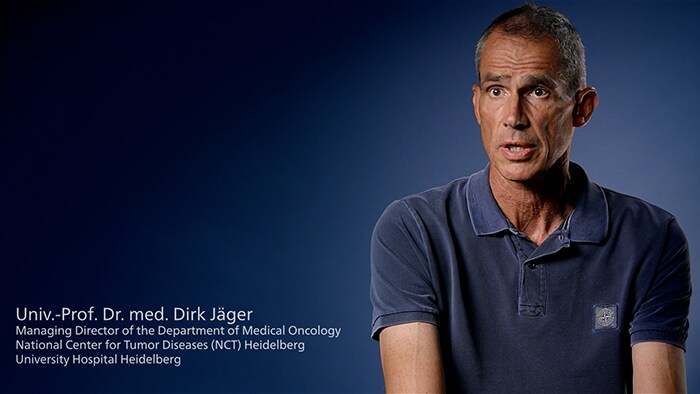
Prof. Dr. Dirk Jäger talks about the greater sensitivity with regard to lesion detection of the Philips Spectral CT 7500.
Expanding opportunities for cardiac imaging with spectral-detector CT
“Unlike oncology, we deal with an organ that is constantly moving, which means that we only have very short imaging windows. This is a special challenge - but one that can be solved thanks to Spectral CT 7500 with its wide detector coverage of 8 cm and the high speed,” says Prof. Dr. Dirk Loßnitzer, senior physician at the university clinic.
“A spectral heart scan takes no longer than a conventional recording, which is a gigantic added value.”
Prof. Dr. Dirk Loßnitzer
"With the Spectral CT 7500, we now have a modality that we can use to obtain complex information in a simple way that is radiation- and contrast medium-neutral for cardiac scanning. A spectral heart scan takes no longer than a conventional recording, which is a gigantic added value.” Thanks to the intelligent algorithms of Spectral Cardiac, they are able to quickly reconstruct the coronary arteries. The Precise Cardiac function also improves the display by correcting for movement artifacts.
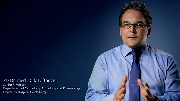
Prof. Dr. Dirk Loßnitzer explores the advantages of the Philips Spectral CT 7500 in cardiac scanning.
Cardiology can now progress from pure coronary imaging to functional, hemodynamically relevant and tissue-characterizing imaging. Heidelberg cardiologists are using Spectral CT 7500 in acute care for examinations of coronary heart disease, congenital and complex heart defects, and also for the planning and control of structural interventions. The system has also proven its worth in helping physicians make diagnoses more easily in challenging clinical heart cases, such as patients with a high, irregular heartbeat or high BMI.
Enhancing the research partnership to improve patient care
Innovations such as Spectral CT 7500 are often the result of joint research work between users and device developers. Heidelberg is looking for quantitative metrics that clinicians can use to make earlier and more reliable diagnostic assessments. Spectral imaging makes it possible to not only significantly increase contrasts for visual assessment, but also to quantify very fine contrasts, for example via quantitative iodine imaging. This is particularly advantageous for oncological imaging. Spectral-detector technology allows assessments of the perfusion status of organs that were previously only possible with more complex techniques. The scientists at Heidelberg hope to use Spectral CT 7500 to obtain more quantitative imaging biomarkers in their research work so that clinicians can bring even more stability and reliability to their diagnostics in the future.
Subscribe to our email updates

Spectral-detector CT scanner
Explore spectral-detector imaging
Footnotes
[1] Analysis by CARTI Cancer Center, Little Rock, AR, USA. Results from Case studies are not predictive of results in other cases. Results in other cases may vary. Disclaimer: Results are specific to the institution where they were obtained and may not reflect the results achievable at other institutions.