• Professional healthcare
  • Products & Services
  • Inspiration
  • Support & Contact
  • Shop
‌
‌
‌
‌
‌
‌
‌
‌
‌
‌
‌
‌
‌
‌
‌
‌
‌
‌
‌
‌
  • Consumer productsConsumer products
    • Home
    • Support
    • Product registration
    • My Philips
  • Healthcare professionalsHealthcare professionals
    • Home
    • Products
    • Services
    • Specialties
    • Solutions
  • Other business solutionsOther business solutions
    • Lighting
    • Automotive
    • Dictation Solutions
    • More
    • Hearing solutions
    • Display solutions
  • About usAbout us
    • Home
    • News
    • Investor Relations
    • Careers
    • More
    • Innovation
    • Environmental, Social and Governance
  • Contact and supportContact and support
    • Customer support
    • Professional healthcare support
    • Company contacts
  • Stay up-to-dateStay up-to-date
  • Select your country / languageGlobal / English
  • Privacy Notice
  • Terms of use
  • Cookie notice
© Koninklijke Philips N.V., 2004 - 2025. Με την επιφύλαξη παντός δικαιώματος.
‌
‌
‌
‌
‌
‌
‌
‌
‌
‌
‌
‌
‌
‌
‌
‌
‌
‌
‌
‌
  1. Image-guided therapy
  2. Devices
  3. Intracardiac echocardiography (ICE) catheter
ICE Catheter
Image-guided therapy

Intracardiac echocardiography (ICE) catheter

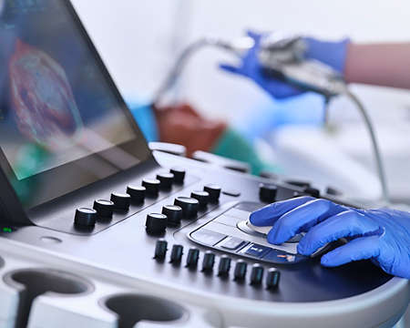
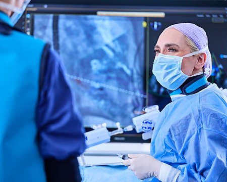
Enhanced image quality for optimal guidance and streamlined workflows

Our innovative intracardiac echocardiography (ICE) solutions elevate the standard of care for electrophysiology and structural heart disease. With clear 2D and 3D imaging our solutions offer an excellent alternative to transesophageal echocardiography (TEE). They provide confidence and control in interventional procedures, while reducing the reliance on general anesthesia.

Image gallery

  • LAAO view via TrueVue 3D rendering and MPRs: 3D imaging mode - Image courtesy: Dr. Andrea Natale, St. David’s Medical Center

    LAAO view via TrueVue 3D rendering and MPRs: 3D imaging mode - Image courtesy: Dr. Andrea Natale, St. David’s Medical Center

  • PFA device in the left pulmonary vein: 2D imaging mode - Image on file with Philips

    PFA device in the left pulmonary vein: 2D imaging mode - Image on file with Philips

  • Assessing position and orientation of TriClip device below leaflets using xPlane: 2D imaging mode - Image on file with Philips

    Assessing position and orientation of TriClip device below leaflets using xPlane: 2D imaging mode - Image on file with Philips

Enabling technologies for Intracardiac echocardiography (ICE) catheter

  • xMatrix transducer technology Thumbnail

    xMatrix transducer technology

    xMATRIX transducer technology enhances image clarity and eases workflow to make exams faster and easier for both clinicians and patients.

Related procedures

  • Ablation and cardiac rhythm management thumbnail

    Ablation and cardiac rhythm management

    Solutions for ablation, cardiac rhythm management and atrial fibrillation to enhance clinical confidence and streamlined workflow in the EP lab.

  • Left atrial appendage occlusion thumbnail

    Left atrial appendage occlusion

    Optimize LAAO procedures with Philips imaging technology, for precise device placement and streamlined workflows.

  • Close to the heart

    Transcatheter mitral and tricuspid therapies

    Elevate transcatheter mitral and tricuspid therapies with Philips imaging for accurate device placement and procedural efficiency.

Frequently asked questions

Transesophageal echocardiography (TEE) and intracardiac echocardiography (ICE) are advanced modalities for cardiac imaging. TEE uses an ultrasound probe in the esophagus near the heart, offering high-resolution images from behind the heart, but requires general anesthesia, which can be uncomfortable. ICE involves placing a catheter inside heart chambers via femoral access, providing direct visualization, near-field imaging and real-time guidance, reducing the need for general anesthesia. Philips provides solutions for both imaging modalities to assist in many clinical areas and patient needs.

Avoidance of TEE and general anesthesia can help improve patient experience:

7-12% of TEE patients experience a procedural injury [4].

40% of TEE patients experience complex lesions (i.e., esophageal hematomas, lacerations, etc.) [5].

By eliminating endotracheal intubation patients are less likely to experience esophageal tears, aspiration pneumonia and stomach lacerations [5,6]. Patients schedule for general anesthesia have statistically higher anxiety scores than regional anesthesia [7].

Moderate sedation is associated with shorter procedure durations and recovery periods [8,9].

While catheter technologies may differ, the terms 4D and 3D refer to the same category of imaging. The JACC Imaging Position Statement on 3D ICE states “the term real time 3D-ICE”[10] is a more accurate nomenclature for the technology given that all echocardiographic modalities are essentially real time."

Philips VeriSight Pro offers 2D and 3D imaging on a single ultrasound platform – the industry-leading Philips EPIQ Ultrasound System. Together, they deliver a comprehensive image guidance solution with options for every need. Compatible EPIQ Systems include: EPIQ 7C, EPIQ CVx and EPIQ CVxi.

We pride ourselves in offering thorough and personalized 3D ICE training industry wide. We offer didactic courses, hands-on simulator training, center of excellence observation, proctorships and more for both physicians and supporting hospital staff. Please contact us to see what training offerings will enable you to reach your program goals.

Footnotes
  1. Diaz JC, Bastidas O, Duque M, et al. Impact of intracardiac echocardiography versus transesophageal echocardiography guidance on left atrial appendage occlusion procedures: A meta‐analysis. J. Cardiovasc. Electrophysiol. 2024; 35(1)44-57.
  2. Amin, AK, Robinson, A, Gundrum E, et al. Intracardiac echocardiography guided left atrial appendage closure contributes to significant cost savings and reduced length of stay. Heart Rhythm. 2019; 16(5) May Supplement: S128.
  3. Dallan LAP et al. Safety, Efficacy, and Cost-Effectiveness of Same-Day Discharge for Left Atrial Appendage Occlusion. J Invasive Cardiol. 2022; 34(2): E124-E131 
  4. Akella K et al. Evaluating the role of transesophageal echocardiography (TEE) or intracardiac echocardiography (ICE) in left atrial appendage occlusion: a meta-analysis. 2021. J Interv Card Electrophysiol. 2021; 60:41-48.
  5. Freitas-Ferraz AB et al. Safety of Transesophageal Echocardiography to Guide Structural Cardiac Interventions. J Am Coll Cardiol. 2020; 75(25):3164-73.
  6. Hemam ME et al. Left atrial appendage closure with the Watchman device using intracardiac vs transesophageal echocardiography: Procedural and cost considerations. Heart Rhythm. 2019; 16:334-342.
  7. Celik et al Evaluation of preoperative anxiety and fear of anesthesia using APAIS score Eur J Med Res (2018) 23:41
  8. Frohlich GM et al. Local versus general anesthesia for transcatheter aortic valve implantation (TAVR) – systematic review and meta-analysis. BMC Medicine. 2014; 12:41.
  9. Wang L et al. Benefit of early discharge among patients with low-risk pulmonary embolism. PLoS ONE. 2017; 12(10):e0185022.
  10. Gilbert H.L. Tang, et al. “Structural Heart Imaging Using 3-Dimensional Intracardiac Echocardiography: JACC Imaging Position Statement.” JACC. Cardiovascular Imaging, 6 June 2024, doi.org/10.1016/j.jcmg.2024.05.012, https://doi.org/10.1016/j.jcmg.2024.05.012. Accessed 13 Aug. 2024.
Disclaimer
Always read the label and follow the directions for use.​
Philips medical devices should only be used by physicians and teams trained in interventional techniques, including training in the use of this device.​ Products subject to country availability. Please contact your local sales representative.​
©2025 Koniklijke Philips N.V. All rights reserved. Trademarks are the property of Koninklijke Philips N.V. or their respective owners. Philips reserves the right to change product specifications without prior notification.